Saturday, 24 October 2015

ACTIVITAT ENZIMÀTICA DE LA CATALASA


  • Introducció:
Valore de forma quantitativa l'activitat enzimàtica de la Catalasa del fetge de pollastre. Dividirem la pràctica en dues parts. En la primera observarem la diferència d'activitat de la catalasa en diferents teixits animals i vegetals. Per aquesta part utilitzarem la patata, tomàquet, pastanaga, cor i el fetge de pollastre. en la segona part veurem la influència de determinats factors en l'activitat enzimàtica.

La catalasa és un enzim present ens els peroxisomes de les cèl·lules animals i vegetals que s'encerrega d'eliminar l'aigua oxigenada que es forma en algunes reacciones del metabolisme. La reacció químia és la següent:

2 H2O2 (l) → 2 H2(l) + O2 (g)

  • Material:
-Diferents teixits animals.
-Patata, tomàquet i pastanaga.
-Tubs d'assaig de coll ample.
-Termòmetre.
-Aigua oxigenada 3%.
-Pinces.
-Tisores.
-Bisturí.

  • Protocol:
Experiment 1: Quin teixit presenta més activitat de la catalasa?

-Tallarem 2 trossos la patata, el tomàquet i pastanaga que pesin més o menys el mateix (1 cm3)
-També tallarem un tros de fetge i cor de la mateixa mida i pes.
-Ho posem en cinc tubs amb 5ml d'aigua destil·lada i 2ml d'aigua oxigenada. I mesurarem l'alçada en mm de les bombolles.
-Patata: 0,7gr, Tomàquet: 0,9, Pastanaga: 0,9, Fetge: 0,7 i Cor: 1gr.


  • Preguntes:
Variable dependent i independent? Dependent: alçada de les bombolles i Independent: els diferents teixits.
Problemes què es vol investigar? Quin teixit presenta més activitat de la catalasa?
Explicació dels resultats: La catalasa presenta més activitat en els teixits animals.

Experiment 2: Com afecten diferents factors en l'activitat de la catalasa?

1er Tub: tros de teixit animal a temperatura ambient (T=19º) (pes: 1gr)
2n Tub: tros de teixit animal amb 10ml d'HCl al 10% (pes=0,89 gr)
3er Tub: tros de teixit animal congelat(T=3º) (pes: 0,7 gr)
4at Tub: Tros de teixit animal bullit (pes: 0,5 gr)
5è Tub: tros teixits submergit en una dissolució saturada de NaCl (pes: 0,7gr)


       *Amb 5ml d'aigua destil·lada i 2ml d'aigua oxigenada.


  • Preguntes:
-Variable dependent i independent? Dependent: alçada de les bombolles, Independent: diferents tractaments.
-Problema què es vol investigar? Com afecten els diferents factos en l'activitat de la catalasa?
-Explicació dels resultats: El teixit que ha estat tractat amb NaCl presenta una activitat de la catalasa.
  • Quina és la funció de la catalasa en els teixits animals i vegetals? On es troba aquest enzim?
Catalitza la descomposició del peròxid d'hidrogen (H202) en oxigen i aigua. A nivell cel·lular es localitza en les mitocòndries i els peroxisomes, excepte en els eritròcits, on es troba en el citosol.

  • Per què quan ens fem una ferida ens posem aigua oxigenada?
Perquè en el cas que hi hagui bacteris, tan aeròbics com anaeròbics, els elimina evitant una possible infecció.


Sunday, 31 May 2015

L21: The chloroplast and the photosynthesis.

  • Material:
-Algae 
-600 ml beaker
-Test tube
-Funnel
-Tap water 
-Light source
-Ruler
 
  • Procedure:
1. First we assigned the different distances to do the experiment and compare the results to each group.
2. We took the 600 ml beaker and placed 7 g of an algae under a clear funnel inside the beaker (the wide end goes over the algae like in the image). The funnel was raised off the bottom on pieces of blue-tack to allow unhampered diffusion of CO2 to Elodea. 
3.We didn't have sodium bicarbonate so we filled the beaker with tap water, the algae and the funnel should be completely under the water.
4. Then we filled a test tube with tap water and placed the thumb over the end of the test tube. We turned the test tube upside down taking care that no air enters and no water comes out and we put this test tube over the end of the funnel (the skinny part)
5. We marked the level of the water on the surface of the test tube with a marker pen.
6. Each group placed the preapartion close to a light source, each group placed the preparation in a different distance 5, 10, 20 and 25 cm, and one with no light source.
7. We also measured the temperature.
8. Finally we left this preparation for and hour and a half. After this time we measured the difference of gas accumulation on the top of the test tube.
 

  • Questions:
1. Identify the dependent and the independent variable of this experiment.
Dependent: gas production, Independent: distance (intensity of the light)
 
2.Using the data from your results prepare a graph and describe what happened to the amount of gas in the test tube.


3.How much gas was producted in the test tube after one hour? And an hour and a half?
I  put the results in the graph.
 
4.Write the photosynthesis equation. Explain each part of the equation. Which subtances are produced by photosynthesis. Which gas is produced that we need in order to live? Plants take in carbon dioxide by diffusion through their stomata. Light energy enters the plant via leaves and water and nutrients enter through roots. The plant is then able to make glucose and oxygen. The glucose moves from the leaves to the plant and the oxygen diffuses out of the leaves. The gas that we need in order to live is oxygen.


L20: Mitosis in an onion root.

  • Material: microscope slide, coverslip, dropper, needles emmanegades, watch glass, beaker, fine tweezers, lancet, encenador and dad filter.
  • Chemicals: aigua destil·lada, aigua corrent, orceïna A i orceïna B.
  • Natural products: onions.
  • Procedure:
  1. Keeps several days a bulb onion on a glass full of water, so that the lower part of bulb where the roots emerge, in contact with water. To hold the onions can be three sticks nailed to utility bulb.
  2. When the roots have grown about three centimeters, cut with scissors and place them end four millimeters in a crystal clock with two millimeters orceïina A.
    Then, heat it with the flame of a lighter until steam appears tenuous. It must ensure that during this process the temperature exceeds 60 ° C at any time.
    To check this, we constantly have to remove the glass clock above the flame and put it over on the back of the hand, which in no case should notice a burning sensation.
  3. With fine tweezers, place one of the pieces of root on a slide and add a few drops of orceine B with dropper.
  4. With the lancet, cut the two millimeters final piece of root and retira'n the rest. Put it on the coverslip and three or four strips of papaya filter. Then make it a slight pressure with the thumb, starting gently and then a little harder, trying not to break the coverslip in order to extend the cells.
  5. Observe it with a microscope 600 increase (or preferably more). As luck you have, you will see cells in different phases of mitotic division.


Tuesday, 7 April 2015

L15: Animal cells vs. Plant cells

  • Objectives:
  1. Identify the major components of cells.
  2. Differetiate between animal and plants cells.
  3. Measure dimensions of the entire cell and the nucleus.


Staining: iodine.


Staining: Methylene blue.

Monday, 23 March 2015

L19: Cells Organelles.


Tomato cromoplast (400x)


Chloroplasts of Vallisneria sp

 
Carrot Cromoplast (100X)


Red Cabage Cloroplast (400X)
Red Cabage Stoma (1000X)
Red Cabage (100X)


L16: Life in a drop of water.

400X


It is a unicellular being, eukaryotic flagellated.


Sunday, 8 March 2015

L17:Gram Staining.

  • Objectives:
  1. Differentive yogurt bacteria.
  2. Relative the staining procedure with the structure of the cells. 

    MAGNIFICATIONS: 400X

Saturday, 31 January 2015

P12: DNA Extraction.

  • Introduction:
Deoxyribonucleic acid (DNA)  is a nuclic acid that encodes the genetic instructions used in the development and fuctioning of all known living organsims and many viruses.
Nucleic acids are biopolymers formed by simple units called nucleotids. Each nucleotide is composed of a nitrogen-containing nucleobase (G, T, C, A) as well as a monosaccharide (deoxyribose) and a phosphate group.
Most DNA molecules consist of two strands coiled around each other to form a double helix. The two strands run in opposite directions to each other and are therefore anti-parallel. Moreover the bases of the two opposite strands unit according to base pairing rules : A-T and G-C.
  • Materials:
  1. 1L Erlenmeyer flask.
  2. 100mL beaker.
  3. 10mL graduated cylinder.
  4. Small funnel.
  5. Glass strining rod.
  6. 10mL pipet.
  7. Knife.
  8. Safety goggles.
  9. Cheesecloth.
  10. Kiwi.
  11. Pineaple juice (1mL/5mL).
  12. Distilled water.
  13. 90% Ethanol ice-cold.
  14. 7mL DNA buffer.
  15. 50mL dish soap.
  16. 15g NaCl.
  17. 900mL tap water.
  • Procedure:
Put the ethanol in the freezer-you will need it really cold later.
Prepare the buffer in a 0,5L beaker: Add 450mL of tap water, 25mL of dish soap and 7g NaCl. Stir the mixture.
  1. Pell the kiwi and chop it to small pieces. Place the pieces of the kiwi in one 600mL beaker and smash with a fork until it becomes a juice puree.
  2. Add 8mL of buffer to the mortar.
  3. Mash the kiwi puree carefully for 1 minutewithout creating many bubbles.
  4. Filter the mixture: put the funnel on top of the graduated cylinder. Place the cheesecloth on top of the funnel. 
  5. Add beaker contain carefully on top of the cheesecloth to fill the graduated cylinder. The juice will drain through the cheesecloth but the chucks of kiwi will not pass through into the graduated cylinder.
  6. Add the pineaple juice to the green juice (you will need about 1mL of pineaple juice to 5mL of the green mixture DNA solution). This step will help us to obtain a purer solution DNA. Pineaple juice contains an enzyme that breaks down proteins.
  7. Tilt the graduated cylinder and pour in an equal amount of ethanol with an automatic pipet. Put the ethanol through the sides of the graduated cylinder very carefully.You will need about equal volumes of DNA solution to ethanol.
  8. Place the graduated cylinder so that it is eye level. Using the stirring rod, collect DNA at the boundary of ethanol and kiwi juice. Do not stir the kiwi juice; only stir in the above ethanol layer!!
  9. The DNA Precipitate looks like long, white and thin fibers.
  10. Gently remove the stirring rod and examine what DNA looks like.
  • Solution: 


  • Questions:
  1. What did the DNA looks like? The DNA looks like long, small white and thin fibers.
  2. Why do you mash the kiwi? Where it is located inside the cells? Because you want to liberate the DNA that is located inside the nucleus.
  3. Explain what is the function of every compund of the buffer (soap ans salt) The salt breaks the nucleus and the cell and the soap takes away the proteins.
  4. DNA is soluble in water, but not in ethanol. What does this fact have to do with our method of extraction? This means that we can only see the DNA in the part of the ethanol beucause if it touches the water it will dissolve.

Sunday, 25 January 2015

P11: Cytochrome C Comparison Lab

  • Introduction:
To compare the relatedness between organisms by examinig the amino acid sequence in the protein, Cytochrome C.
Cytochrome C is associated with the inmer membrane of the mitochondrion. It is a small protein from eucariote cell. It is a Hemeprotein; iron group.
Function: produce energy, is a part of a electron transport chain (ATP).
  • Procedure:
We compare the mammals: horse, donkey, whale; beardds: penguin, chicken; insect: moth and fungui: wheat, yeast.
  • Conclutions:


P10: Protein Denaturation I

  • Introduction:
Denaturation is a process in which proteins or nucleic acids lose the quaternary, tertiary and secondary structure that is present in their native state. Denaturation is the result of the application of some external stress (heat and pH change) or compounds such as a strong acid or base, aconcentrated inorganic salt or organic solvent. In very few cases denaturation is reversible and proteins can recuperate their native state when the denaturing factor is removed. This process is called renaturation.
  • Materials:
-2x250mL beaker.
-4 test tubes.
-Test tube rack.
-10 mL pipet.
-Knife.
-Glass marking pen.
-Potato.
-Distilled water.
-Hydrogen Peroxide.
-NaCl.
-HCl.
  • Catalase Activity:
Catalase is a common enzye found in nearly all-living organisms exposed to oxygen. It catalyzes the decomposition of hydrogen peroxide (H2O2) to water and oxygen. It is a very important enzyme in protecting the cell from oxidative damage  and preveting the accumulation of hydrogen peroxide.


                                             2 H2O2 --------------------->   2 H2O + O

Catalase is a tetramer of four polypeptide chains, ecah over 500 amino acids long. It contains four Porphyrin Heme groups (iron groups) that allow the enzyme yo react with the hydrogen peroxide. The optimum pH for human catalase is aprox. 7, in other organisms vary between 4 and 11. The organelle that stores catalase in eukaryotic cells is the peroxisome, which also contains peroxidases.

  • Procedure:
In this experiment we aregoing to test the catalase activity in different enviroment situations. we are measures the rate of enzyme activity under varios conditions, such as different pH values and temperature. We will measure catalase activity by observing the oxygen gas bubbles when H2O2  is destroyed. If lots of bubbles are produced, it means the reaction is happening quickly and the catalaseenzyme is very active.
  1. Prepare 30mL of H2O2  10% in a beaker (use a pipet).
  2. Prepare 30mL of HCl 10% in a beaker.
  3. Prepare 30mL of NaCl 50% in a beaker.
  4. Peel a fresh potato tuber and cut the tissue in five cubes of  1cm3. Weigh them and equal the mass.
  5. Label 5 test tubes (1,2,3,4,5).
  6. Immerse 10 minutes your piece of potato inside HCl beaker.
  7. Immerse 10 minutes another piece of potato NaOH beaker.
  8. Boil another piece of potato.
  9. With a mortar, mash up the third piece of potato.
  10. Add 5mL H2O2  10% in each test tube.
  11. With a glass-marking pen mark the height of the height of the bubbles.
  12. Compare the results of the test 5 test tubes.
  • Conclutions:
Mash potato has more activity because proteins are more exposed and react more.
The raw potato is the second having more activity because it has not had any previous alteration.
Potato with NaCl practically no present activity.
 Boiled potato and potato with HCl no present activity because the proteins were denatured and above.
-Completed the table below with the important parts of this experiment:
Parts:
In this experiment this was...
Independent variable
Tratament of each potato.
Dependent variable
The height of the bubbles.
Experimental Group(s)
Boiled, with HCl, with NaCl and mashed up potato.
Control Groups
Raw potato.
Constants
Weight, same amount of H2O2 , time...
-Represent your results in a chart: Treatment vs bubble's height.

  • Question:
  1. How did the temperature of the potato affect the activity of catalase?
  2. How did the change of the pH of the potato affect the activity of catalase?
  3. In which potato treatment was catalase the most active? Why do you think this was?


Sunday, 11 January 2015

SEVENTH EXPERIMENT: Protein Identification

  • Introduction:
Biuret's test is a chemical test used for detecting the presence of peptide bonds. Polypeptides as proteins, are chains of amino acids link together by peptide bonds.
The biuret reaction can be used to assess the concentration of proteins because peptide bonds occurs with the same  frequency per amino acid peptide.
  • Materials:
-7 × 250mL beaker.
-6 test tubes.
-Test tube rack.
-6 × 10mL Pipet.
-Mortar.
-Glass marking pen.
-Gloves.
-Goggles.
-Milk.
-Rice Milk.
-Egg.
-Yogurt.
-Potato.
-Distilled water.
-NaOH 20%.
-10 drops of CuSO4.
  • Procedure:
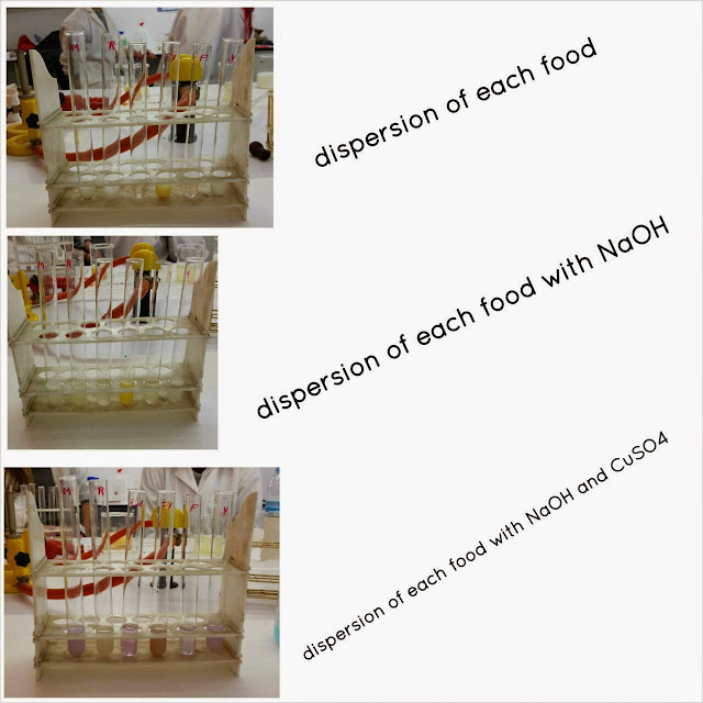
We are going to determine protein protein concentration in some foods: milk, rice milk, yogurt, potato and egg. Be careful, aa we are going to test separately the egg white and the yolk. Put gloves and use goggles, remember that NaOH is caustic!!
First of all we are going to dilute the protein.
1.  Add 100mL of distilled water to each 250mL beaker. Label them with M(milk), R(rice milk) and EW(egg white), EY(yolk) and P(potato).
2. Separate the egg white and the yolk in another beaker.
3. Smash the potato in a mortar and add some amount of the of the smashed potato to the P beaker.
Prepare the samples:
4. Add 10mL of a dispersions of each food (M, S, EY, EW and Y) to the indicate beaker. Calculate the final concentration. All the groups will use the same dispersion from the beakers.
5. Prepare 6 test tubes (clean and dry) and label M, S, EW, EY, Y and P. Add 2mL of the every food dilution of each beaker.
6. Add 2mL of 20%NaOH dissolution.
7. Shake gently and add 5 drops of CuSO4 in each tube. Allow the mixture to stand for 5 minuts.
8. Note any colour change. Remember that proteins will turn solution pink or purple.
9. Compare the test tubes.
  • Questions:
1. Wich food has proteins? Egg white, milk, yogurt, potato.
2. Wich food has more proteins? Why? Egg white, because they are animal food and is rich in proteins.
3. Do you find any difference between soy rice and cow milk? Yes, rices milk doesn't have proteins, however cow milk does.
4. Is there any difference among milk and yogurt? Why? Yes, because yogurt has more proteins than milk.